Skip to content
  • Newsletter
  • About us
  • Contact
  • EN
  • DE
  • Newsletter
  • About us
  • Contact
  • EN
  • DE
Search
Close
  • Overview
  • Research
  • Here & now
  • Smart minds
Menu
  • Overview
  • Research
  • Here & now
  • Smart minds
  • Overview
  • Research
  • Here & now
  • Smart minds
  • Contact
  • Newsletter
  • About us
Menu
  • Overview
  • Research
  • Here & now
  • Smart minds
  • Contact
  • Newsletter
  • About us
Search
  • EN
  • DE
10. April 2017

Metal under the microscope: IW shows prizewinning pictures

IW | “Different appearances of metal“: This is the title of a photo exhibition in East Friesland, Germany, organized by the Institute of Materials Science (IW) of Leibniz Universität Hannover. The pictures have been created by five employees working in the metallography sector of the Hannover Centre for Production Technology (PZH).

Just few people are aware of the unique look of metal under a light microscope. This is why the Institute of Materials Science (IW) has organized a photo exhibition which gives a microscopic insight into the structure of metals. 
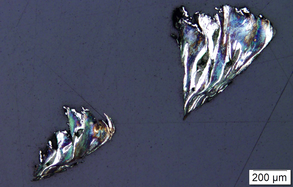
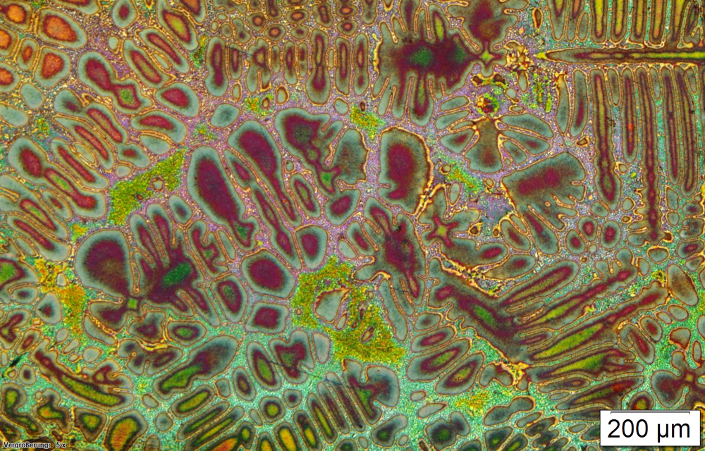
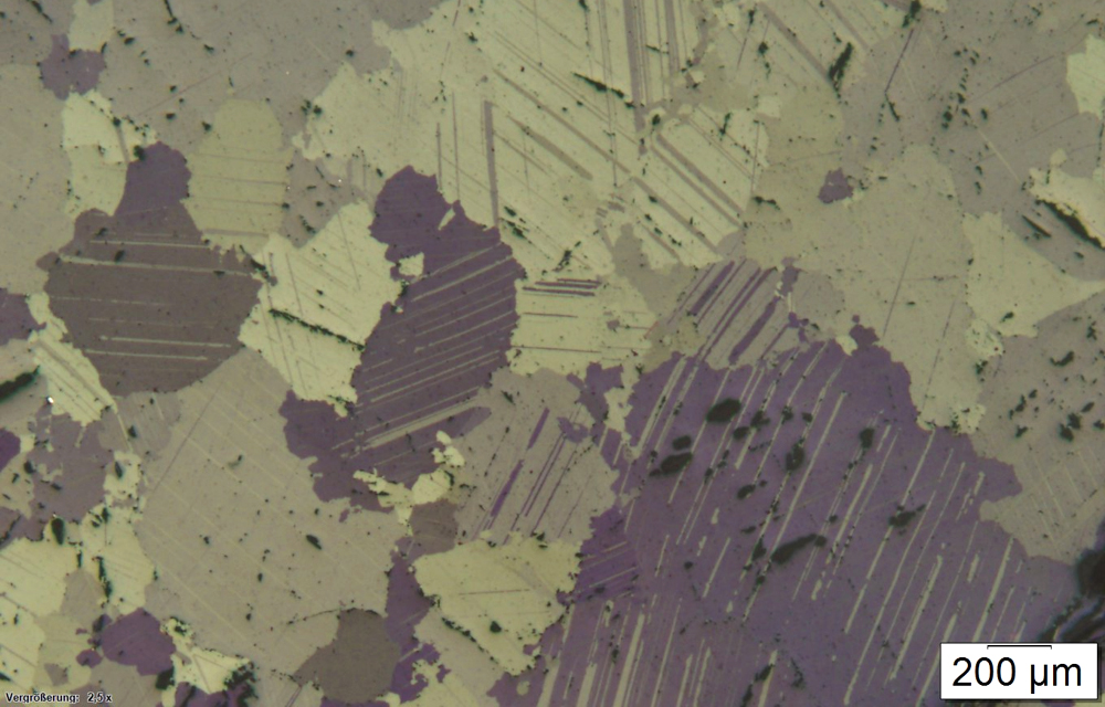
The structures are visualized by microscopic images of metal samples, so-called micrographs. Depending on the material and purpose of investigation, they are ground, polished and sometimes treated with various etching agents. Micrographs provide materials scientists important insights, because microscopic images are able to visualize interactions and processes in the material, as they are caused by temperature influences or deformations. 
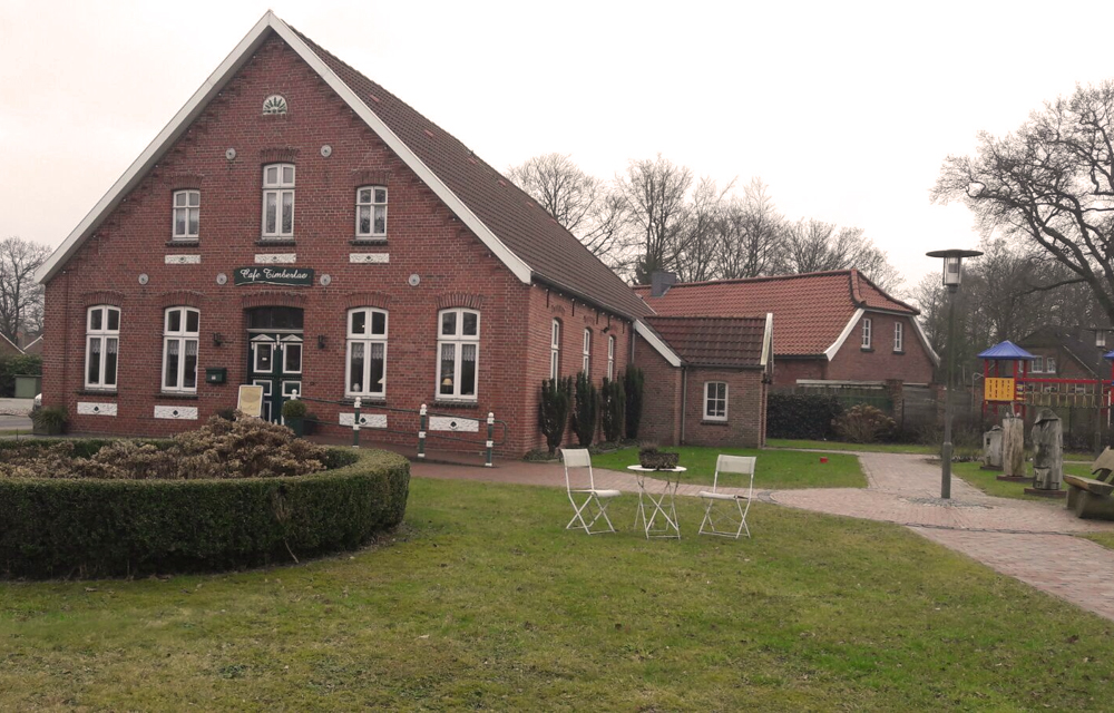
The pictures have already won several prizes. Since 10 April, they can be seen for three months at the Café Timberlae (Leerer Landstraße 12, Großefehn/Timmel, Germany). 

by Judith Kebbe

Similar posts

  • Newly awarded: Metallography in materials science

At a glance

  • Photo exhibition of metals under the microscope
  • Café Timberlae, Leerer Landstraße 12, Großefehn/Timmel, Germany
  • 10 April to 30 June, daily from 11 a.m. to 18 p.m.
IW_Ausstellung_Bild1_02
Bright metal under the light microscope: For this image steel chips were etched with nitric acid. (Photo: Mareike Reiter)
IW_Ausstellung_Bild2_01
This microscopic image shows cast aluminium which was colour-etched with Weck’s reagent. (Photo: Ute Teuber)
IW_Ausstellung_Bild3_01
Different appearances of unetched marble under a light microscope. (Photo: Mareike Reiter)
IW_Ausstellung_Bild4_01
East Frisian insider tip: Café Timberlae in Großefehn/Timmel. (Photo: Ines Knälmann)
Previous
Next

Contact

Mareike Reiter

0511 762-18051

reiter@iw.uni-hannover.de

www.iw.uni-hannover.de

This Page

drucken

recommend

  • tweet 

Similar posts

  • Newly awarded: Metallography in materials science

This Page

drucken

recommend

  • tweet 

ISSN 2198-1922 | Legal Information | Privacy notice | Article Sitemap